Collagen Structure & Function: Difference between revisions
No edit summary |
Luis Netto (talk | contribs) No edit summary |
||
| (37 intermediate revisions by 3 users not shown) | |||
| Line 1: | Line 1: | ||
< | <StructureSection load='1cag' size='450' side='right' scene='Sandbox_168/Default/3' caption=''> | ||
==Introduction== | |||
[[Collagen]] is a member of a family of naturally occurring proteins. It is one of the most plentiful proteins present in mammals and it is responsible for performing a variety of important biological functions. It is most well-known for the structural role it plays in the body. It is present in large quantities in connective tissue and provides tendons and ligaments with tensile strength and skin with elasticity. It often works in conjuction with other important proteins such as keratin and elastin. | |||
== | ==Biosynthesis== | ||
Collagen synthesis begins specialized cells called fibroblasts <ref name="biosyn">PMID:PMC1367617</ref>. It is here that amino acids undergo activation; Proline is hydroxylated to Hydroxyproline and Lysine to Hydroxylysine. Peptide subunits of ~250 residues are assembled on the ribosome and are linked by carbohydrate residues to form α-chains <ref name="biosyn" />. Three α-chains then associate with each other and then further associate extracelluarly forming a molecule with a molecular weight of 360,000 <ref name="biosyn" />. Bonds are further strengthened thus forming the insoluble collagen fibril <ref name="biosyn" /> <ref name="collalike" />. During the process of collagen synthesis, free-hydroxyproline and hydroxylysine peptides appear as by-products, some of which are metabolized and may appear in urine <ref name="biosyn" />. | |||
==Molecular Structure== | ==Molecular Structure== | ||
The shape and structural properties of a native collagen molecule are established by its triple-helical domain(s). In classical collagen molecules a single triple-helical domain is observed to compose close to 95% of the molecule <ref>PMID: 19853297</ref>. However there are also other types of collagens that have been shown to comprise of multiple triple-helical domains which only account for a fraction of the molecule's overall mass. | The shape and structural properties of a native collagen molecule are established by its triple-helical α-domain(s). In classical collagen molecules a single triple-helical domain is observed to compose close to 95% of the molecule.<ref name="residues">PMID:19853297</ref>. However there are also other types of collagens that exist which have been shown to comprise of multiple triple-helical α-domains which only account for a fraction of the molecule's overall mass. | ||
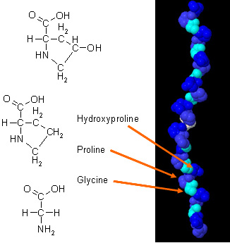
The triple-helical domain structure of collagens consists of three distinct α-chains and earns collagen the name "tropocollagen" <ref name="collalike">PMID:7695699</ref>. Each of these chains contain a characteristic L-handed amino acid sequence of polyproline, often termed as polyproline type II helix <ref>PMID: 19344236</ref>. The proper folding of each of these chains requires a glycine residue to be present in every third position in the polypeptide chain. For example, each α-chain is composed of multiple triplet sequences of of Gly-Y-Z in which Y and Z can be any amino acid. Y is commonly found as proline and Z is usually present as hydroxyproline (Figure 1.). The presence of hydroxyproline in the Y position is also thought to contribute to the stability of the helical form <ref name="collalike" />. | |||
The image on the right-hand side has each side chain colored a different color to shown how each individual <scene name='Sandbox_168/Helices/1'> | These three α-chains are then twisted around one another in a rope-like manner to produce the overall tightly packed triple-helical form of the molecule. The interaction of α-chains is stabilized via interchain hydrogen bonding making the molecule fairly resistant to attack by other molcules. Each α-chain is surrounded by a hydration sphere which allows a hydrogen bonding network to be present between the water molecules and the peptide acceptor groups.<ref name="collalike" />. This hydrogen bonding occurs when the amino group (NH) of a glycine residue forms a peptide bond with the carbonyl (C=0) of an adjacent residue. The overall molecule is approxiametly 300nm long and 1.5-2nm in diameter.<ref name="collalike" />. | ||
The image on the right-hand side has each side chain colored a different color to shown how each individual <scene name='Sandbox_168/Helices/1'>helices</scene> interact with the others to form the overall molecule. The <scene name='Sandbox_168/Myscene/1'>active sites</scene> | |||
have also been illustrated to point out their positions in the triple-helix. | have also been illustrated to point out their positions in the triple-helix. | ||
[[Image:collagen_(alpha_chain).jpg | thumb | Amino Acid residues in collagen. | [[Image:collagen_(alpha_chain).jpg |400px| thumb |'''Figure 1.''' Amino Acid residues in collagen. Gly, Pro and Hydroxyproline residues present in a collagen molecule <ref name="residues"/>.]] | ||
{{Clear}} | |||
==Function== | ==Function== | ||
There are | There are close to 30 different types of collagen that have been identified so far.<ref name="types">PMID:17581806</ref>. | ||
The most abundant type of collagen present in the human body is that of Type I <ref name="types" /> with significant amounts of Type II,III and IV also accounted for. | |||
*Collagen I- found in bones,tendons,organs | *Collagen I- found in bones,tendons,organs | ||
| Line 27: | Line 27: | ||
*Collagen III- found mainly in reticular fibres | *Collagen III- found mainly in reticular fibres | ||
*Collagen IV- found in the basement membrane of cell membranes | *Collagen IV- found in the basement membrane of cell membranes | ||
*Collagen V- found in hair | *Collagen V- found in hair,nails | ||
==Collagen-Related Disorders== | ==Collagen-Related Disorders== | ||
There are many types of disorders associated with collagen.These disorders typically occur as a result of improper folding of these molecules and occasionally due to a particular amino acid substitution | There are many types of disorders associated with collagen.These disorders typically occur as a result of improper folding of these molecules and occasionally due to a particular amino acid substitution <ref name="collalike" />. These include: | ||
*Elhers-Danlos Syndrome (IV) | *Elhers-Danlos Syndrome (IV) | ||
*Alport Syndrome (IV) | *Alport Syndrome (IV) | ||
*Osteogenesis imperfecta (I) | *Osteogenesis imperfecta (I) | ||
-more commonly known as Brittle Bone disease | |||
*Chondrodysplasias (II) | *Chondrodysplasias (II) | ||
*Atopic Dermatitis (III) | *Atopic Dermatitis (III) | ||
</StructureSection> | |||
==References== | ==References== | ||
<references/> | <references/> | ||
Latest revision as of 16:15, 19 May 2018
IntroductionCollagen is a member of a family of naturally occurring proteins. It is one of the most plentiful proteins present in mammals and it is responsible for performing a variety of important biological functions. It is most well-known for the structural role it plays in the body. It is present in large quantities in connective tissue and provides tendons and ligaments with tensile strength and skin with elasticity. It often works in conjuction with other important proteins such as keratin and elastin. BiosynthesisCollagen synthesis begins specialized cells called fibroblasts [1]. It is here that amino acids undergo activation; Proline is hydroxylated to Hydroxyproline and Lysine to Hydroxylysine. Peptide subunits of ~250 residues are assembled on the ribosome and are linked by carbohydrate residues to form α-chains [1]. Three α-chains then associate with each other and then further associate extracelluarly forming a molecule with a molecular weight of 360,000 [1]. Bonds are further strengthened thus forming the insoluble collagen fibril [1] [2]. During the process of collagen synthesis, free-hydroxyproline and hydroxylysine peptides appear as by-products, some of which are metabolized and may appear in urine [1]. Molecular StructureThe shape and structural properties of a native collagen molecule are established by its triple-helical α-domain(s). In classical collagen molecules a single triple-helical domain is observed to compose close to 95% of the molecule.[3]. However there are also other types of collagens that exist which have been shown to comprise of multiple triple-helical α-domains which only account for a fraction of the molecule's overall mass.
The triple-helical domain structure of collagens consists of three distinct α-chains and earns collagen the name "tropocollagen" [2]. Each of these chains contain a characteristic L-handed amino acid sequence of polyproline, often termed as polyproline type II helix [4]. The proper folding of each of these chains requires a glycine residue to be present in every third position in the polypeptide chain. For example, each α-chain is composed of multiple triplet sequences of of Gly-Y-Z in which Y and Z can be any amino acid. Y is commonly found as proline and Z is usually present as hydroxyproline (Figure 1.). The presence of hydroxyproline in the Y position is also thought to contribute to the stability of the helical form [2].
These three α-chains are then twisted around one another in a rope-like manner to produce the overall tightly packed triple-helical form of the molecule. The interaction of α-chains is stabilized via interchain hydrogen bonding making the molecule fairly resistant to attack by other molcules. Each α-chain is surrounded by a hydration sphere which allows a hydrogen bonding network to be present between the water molecules and the peptide acceptor groups.[2]. This hydrogen bonding occurs when the amino group (NH) of a glycine residue forms a peptide bond with the carbonyl (C=0) of an adjacent residue. The overall molecule is approxiametly 300nm long and 1.5-2nm in diameter.[2]. The image on the right-hand side has each side chain colored a different color to shown how each individual interact with the others to form the overall molecule. The have also been illustrated to point out their positions in the triple-helix.  FunctionThere are close to 30 different types of collagen that have been identified so far.[5]. The most abundant type of collagen present in the human body is that of Type I [5] with significant amounts of Type II,III and IV also accounted for.
Collagen-Related DisordersThere are many types of disorders associated with collagen.These disorders typically occur as a result of improper folding of these molecules and occasionally due to a particular amino acid substitution [2]. These include:
-more commonly known as Brittle Bone disease
|
| ||||||||||
ReferencesReferences
- ↑ 1.0 1.1 1.2 1.3 1.4 PMID:PMC1367617
- ↑ 2.0 2.1 2.2 2.3 2.4 2.5 Bella J, Eaton M, Brodsky B, Berman HM. Crystal and molecular structure of a collagen-like peptide at 1.9 A resolution. Science. 1994 Oct 7;266(5182):75-81. PMID:7695699
- ↑ 3.0 3.1 Yamazaki CM, Kadoya Y, Hozumi K, Okano-Kosugi H, Asada S, Kitagawa K, Nomizu M, Koide T. A collagen-mimetic triple helical supramolecule that evokes integrin-dependent cell responses. Biomaterials. 2010 Mar;31(7):1925-34. Epub 2009 Oct 22. PMID:19853297 doi:10.1016/j.biomaterials.2009.10.014
- ↑ Shoulders MD, Raines RT. Collagen structure and stability. Annu Rev Biochem. 2009;78:929-58. PMID:19344236 doi:10.1146/annurev.biochem.77.032207.120833
- ↑ 5.0 5.1 Koide T. Designed triple-helical peptides as tools for collagen biochemistry and matrix engineering. Philos Trans R Soc Lond B Biol Sci. 2007 Aug 29;362(1484):1281-91. PMID:17581806 doi:10.1098/rstb.2007.2115