User:Jing Liu/Sandbox 1: Difference between revisions
New page: frame|Bacterial chemotaxis receptor One of the CBI Molecules being studied in the [http://www.umass.edu/cbi/ University of Massachusetts Amherst Ch... |
No edit summary |
||
| (42 intermediate revisions by the same user not shown) | |||
| Line 1: | Line 1: | ||
[[ | One of the [[CBI Molecules]] being studied in the [http://www.umass.edu/cbi/ University of Massachusetts Amherst Chemistry-Biology Interface Program] at UMass Amherst and on display at the [http://www.molecularplayground.org/ Molecular Playground]. | ||
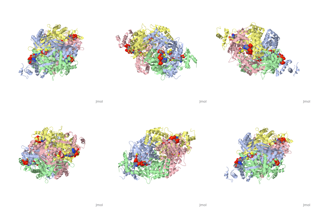
==Your Heading Here (maybe something like 'Structure')==<StructureSection load='1dq8' size='300' side='right' caption='Structure of HMG-CoA reductase (PDB entry [[1dq8]])' scene=''>Anything in this section will appear adjacent to the 3D structure and will be scrollable. | |||
1 | |||
2 | |||
2 | |||
3 | |||
3 | |||
4 | |||
5 | |||
5 | |||
6 | |||
6 | |||
7 | |||
8 | |||
8 | |||
9 | |||
10 | |||
11 | |||
12 | |||
</StructureSection> | |||
<Structure load='1L8Q' size='300' frame='true' align='right' caption='1L8Q 76-399' scene='User:Jing_Liu/Sandbox_1/1/4' /> | |||
This is a <scene name='User:Jing_Liu/Sandbox_1/1/4'>DnaA monomer strucure(residues 76-399)</scene>, which contains an <scene name='User:Jing_Liu/Sandbox_1/1/5'>ADP</scene> in it. | |||
---- | |||
---- | |||
---- | |||
=== DnaA function in Prokaryotes=== | |||
DnaA is an AAA+ protein in prokaryotes for DNA replication initiation. To ensure correct timing of DNA replication, intracellular DnaA is under multi-level control, including changing expression/degradation level, regulator control and functional activation/inactivation by nucleotide-depedent conformational change. DnaA is a conserved protein in prokaryotes. In ''E. coli'', There are four regions in this protein[http://www.ncbi.nlm.nih.gov/pubmed/10572294]: | |||
I: residues 1-85. DnaB loading domain; | |||
II: residues 86-133. linker region; | |||
III: residues 134-372. ATPase domain; | |||
IV: residues 373-467. DNA binding domain; | |||
DnaA function is regulated by different nucleotide binding state. In ATP bound state, DnaA can recognize more binding elements on the chromosome(called DnaA box), and initiate replication by unwinding specific region in the DNA and recruit DnaB helicase. However, after the ATP hydrolysis, DnaA-ADP becomes an inactive protein. This activity change was shown to be associated with its oligomeric assembly state. | |||
=== DnaA Monomer Structure === | |||
The <scene name='User:Jing_Liu/Sandbox_1/1/4'>structure of DnaA</scene> in ''Aquifex aeolicus'' was solved in 2002[http://www.ncbi.nlm.nih.gov/pubmed/12234917?dopt=Abstract], and it contains the linker region, AAA+ domain and DNA binding domain(DBD). The interaction with DNA is through the recognization of a conserved 9 bp DNA recognition sequence (TTA/TTNCACC), and this was shown by the crystal structure of the <scene name='User:Jing_Liu/Sandbox_1/2/1'>complex</scene> in ''Mycobacterium tuberculosis''[http://www.ncbi.nlm.nih.gov/pubmed/21620858?dopt=Abstract]. | |||
=== Distinct Assembly State of DnaA === | |||
< | <Structure load='1L8Q' size='300' frame='true' align='right' caption='2HCB tetramer' scene='User:Jing_Liu/Sandbox_1/3/3' /> | ||
caption=' | The crystal structure of AMP-PCP-bound DnaA reveals a <scene name='User:Jing_Liu/Sandbox_1/3/3'>right-handed superhelix</scene> structure[http://www.ncbi.nlm.nih.gov/pubmed/16829961?dopt=Abstract]. Each monomer contact with another two monomers to form a filamentous structure. The engagement of ATP involved the arginine figure at position 230. | ||
=== | === Reference === | ||
1. Messer, W., et al., Functional domains of DnaA proteins. Biochimie, 1999. 81(8-9): p. 819-25. | |||
2. Erzberger, J.P., et al., The structure of bacterial DnaA: implications for general mechanisms underlying DNA replication initiation. EMBO J. 2002 Sep 16;21(18):4763-73. | |||
3. Tsodikov OV and Biswas T. Structural and thermodynamic signatures of DNA recognition by Mycobacterium tuberculosis DnaA. J Mol Biol. 2011 Jul 15;410(3):461-76. Epub 2011 May 18. | |||
4. Erzberger, J.P., et al., Structural basis for ATP-dependent DnaA assembly and replication-origin remodeling. Nat Struct Mol Biol. 2006 Aug;13(8):676-83. Epub 2006 Jul 9. | |||
Latest revision as of 19:01, 10 October 2012
One of the CBI Molecules being studied in the University of Massachusetts Amherst Chemistry-Biology Interface Program at UMass Amherst and on display at the Molecular Playground.
==Your Heading Here (maybe something like 'Structure')==
Anything in this section will appear adjacent to the 3D structure and will be scrollable. 1 2 2 3
3
4 5 5 6 6 7 8 8 9
10 11 12
|
| ||||||||||
|
This is a , which contains an in it.
DnaA function in ProkaryotesDnaA function in Prokaryotes
DnaA is an AAA+ protein in prokaryotes for DNA replication initiation. To ensure correct timing of DNA replication, intracellular DnaA is under multi-level control, including changing expression/degradation level, regulator control and functional activation/inactivation by nucleotide-depedent conformational change. DnaA is a conserved protein in prokaryotes. In E. coli, There are four regions in this protein[1]:
I: residues 1-85. DnaB loading domain;
II: residues 86-133. linker region;
III: residues 134-372. ATPase domain;
IV: residues 373-467. DNA binding domain;
DnaA function is regulated by different nucleotide binding state. In ATP bound state, DnaA can recognize more binding elements on the chromosome(called DnaA box), and initiate replication by unwinding specific region in the DNA and recruit DnaB helicase. However, after the ATP hydrolysis, DnaA-ADP becomes an inactive protein. This activity change was shown to be associated with its oligomeric assembly state.
DnaA Monomer StructureDnaA Monomer Structure
The in Aquifex aeolicus was solved in 2002[2], and it contains the linker region, AAA+ domain and DNA binding domain(DBD). The interaction with DNA is through the recognization of a conserved 9 bp DNA recognition sequence (TTA/TTNCACC), and this was shown by the crystal structure of the in Mycobacterium tuberculosis[3].
Distinct Assembly State of DnaADistinct Assembly State of DnaA
|
The crystal structure of AMP-PCP-bound DnaA reveals a structure[4]. Each monomer contact with another two monomers to form a filamentous structure. The engagement of ATP involved the arginine figure at position 230.
ReferenceReference
1. Messer, W., et al., Functional domains of DnaA proteins. Biochimie, 1999. 81(8-9): p. 819-25.
2. Erzberger, J.P., et al., The structure of bacterial DnaA: implications for general mechanisms underlying DNA replication initiation. EMBO J. 2002 Sep 16;21(18):4763-73.
3. Tsodikov OV and Biswas T. Structural and thermodynamic signatures of DNA recognition by Mycobacterium tuberculosis DnaA. J Mol Biol. 2011 Jul 15;410(3):461-76. Epub 2011 May 18.
4. Erzberger, J.P., et al., Structural basis for ATP-dependent DnaA assembly and replication-origin remodeling. Nat Struct Mol Biol. 2006 Aug;13(8):676-83. Epub 2006 Jul 9.
